
Nuclear Medicine Imaging: Check How Organs Are Functioning Inside Body
Nuclear medicine imaging is a medical technique that uses small amounts of radioactive materials to diagnose and evaluate various diseases by capturing images of how these substances interact with the body's tissues and organs.
What is Nuclear Medicine Imaging
Nuclear medicine imaging is a medical imaging technique.
- It involves the use of small amounts of radioactive materials.
- These materials are called radiotracers or radiopharmaceuticals.
- Radiotracers are injected, inhaled, or swallowed by patients.
- Special cameras detect the radiation emitted by these tracers.
- This helps create images of how organs and tissues function.
- It's used to diagnose and monitor various medical conditions.
Nuclear Medicine Imaging
Here are the basic details for the Nuclear Medicine Imaging .
| Also Known As | Nuclear Scan |
| Type | Diagnostic Imaging |
| Purpose | Assess organ function, detect diseases |
| Preparation | May require specific dietary instructions |
| Fasting | Sometimes fasting is necessary |
| Gender | All genders |
| Age Group | All age groups |
| Procedure Duration | 1-2 hours or longer, depending on the scan |
| Reporting Time | Typically within a few days |
| Cost | 450-900* INR |
| Pregnancy Consideration | Avoided during pregnancy |
| Risks and Safety | Low radiation exposure, generally safe |
| Accessibility | Available in specialized medical centers |
*Price range may vary as per location, facility, type, and procedure.
What are the Purpose or Reasons for Nuclear Medicine Imaging?
Here are common reasons for Nuclear Medicine Imaging.
- Assess organ function and physiology
- Detect and diagnose diseases like cancer
- Visualize bone health andentify fractures
- Evaluate thyroid and other glandular conditions
- Monitor blood flow and circulation
- Locate and analyze tumors or abnormalities
- Guide treatment planning for certain conditions
Types of Nuclear Medicine Imaging
Here are the types of Nuclear Medicine Imaging along with their primary use.
| Nuclear Medicine Imaging | Organ/System | Primary Use |
|---|---|---|
| Bone Scan | Skeleton | Detect bone abnormalities |
| Thyroid Scan | Thyroid gland | Assess thyroid function |
| Cardiac Perfusion Scan | Heart | Evaluate cardiac blood flow |
| PET/CT Scan | Various | Detect metabolic activity |
| Gallium Scan | Whole body | Detect inflammation or tumors |
| Renal Scan | Kidneys | Evaluate kidney function |
| Hepatobiliary Scan | Liver/Gallbladder | Assess liver and bile ducts |
| Lung Ventilation/Perfusion Scan | Lungs | Assess lung function |
| Octreotide Scan | Various | Detect neuroendocrine tumors |
These nuclear medicine imaging types provide valuable insights into specific organs and conditions.
Preparing for Your Nuclear Medicine Imaging: Tips and Information
Here is the basic preparation before, during, and after Nuclear Medicine Imaging for any patient.
Before Nuclear Medicine Imaging:
- Consultation: Schedule the nuclear medicine imaging and discuss any concerns or medical history with your healthcare provider.
- Fasting: Follow any specific fasting instructions provided by your healthcare team if required for your specific scan.
- Medications: Inform your healthcare provider about all medications you are taking, as some may need to be adjusted or temporarily stopped.
- Allergies: If you have known allergies to any medications or substances used in nuclear medicine, inform your healthcare provider beforehand.
- Clothing: Wear comfortable clothing, and you may be asked to change into a hospital gown depending on the area being scanned.
During Nuclear Medicine Imaging:
- Injection: If necessary, you may receive an injection of a radioactive tracer. It's typically administered through an IV or ingested orally.
- Waiting Period: After receiving the tracer, there may be a waiting period for it to distribute within your body before the actual scan.
- Positioning: You will be positioned on the imaging table, and the scanner will be placed over the area of interest.
- Communication: You can communicate with the technician if you have any concerns or discomfort during the procedure.
After Nuclear Medicine Imaging:
- Recovery: There is typically no special recovery needed. You can resume normal activities unless otherwise advised.
- Hydration: Drinking fluids can help eliminate the tracer from your body. Follow any hydration recommendations from your healthcare team.
- Results: Your nuclear medicine imaging results will be reviewed by a radiologist or nuclear medicine specialist, and a report will be sent to your healthcare provider.
- Follow-Up: Schedule a follow-up appointment with your healthcare provider to discuss the imaging results and any necessary next steps or treatments.
Please note that the specific instructions and procedures can vary based on the type of nuclear medicine scan you are undergoing and your individual medical condition. Always follow the guidance provided by your healthcare team for a successful and safe nuclear medicine imaging procedure.
Who Performs a Nuclear Medicine Imaging?
| Professional | Role |
|---|---|
| Nuclear Medicine Technologist | Administers radioactive substances, operates imaging equipment. |
| Radiologic Technologist | Assists during the procedure and helps with patient positioning. |
| Radiologist | Interprets nuclear medicine images, provides a diagnosis. |
Nuclear Medicine Imaging Procedure
The procedure for Nuclear Medicine Imaging typically follows these steps:
- Check-in and registration at the nuclear medicine department.
- You may be asked to change into a gown provided by the facility.
- A radioactive tracer is administered through an injection, oral ingestion, or inhalation.
- You wait for a specific amount of time to allow the tracer to distribute in your body.
- You will lie down on an examination table.
- A gamma camera or PET scanner is used to capture images of the tracer's distribution.
- Multiple scans may be taken from different angles.
- The procedure is generally painless and can take anywhere from 30 minutes to several hours.
- After imaging, you're typically free to leave.
- The images are interpreted by a radiologist or nuclear medicine physician.
- You may receive the results during your visit or at a later time.
Nuclear Medicine Imaging Results
Here are some common elements you might find in a Nuclear Medicine Imaging report:
| Nuclear Medicine Imaging Findings | Interpretation |
|---|---|
| Procedure (e.g., PET, SPECT, etc.) | Description of the imaging technique used |
| Radiotracer Uptake | Areas of increased or decreased radiotracer uptake |
| Specific Area or Organ | Location or organ system being assessed |
| Quantitative Measurements | Quantitative values, if applicable (e.g., standardized uptake values in PET) |
| Functional Information | Assessment of metabolic or functional activity |
| Impression | Summary of key findings or diagnostic impressions |
| Recommendations | Follow-up tests, treatments, or further evaluation, if necessary |
| Conclusion | Final remarks or clinical recommendations |
Please note that the format and content of nuclear medicine imaging reports can vary depending on the specific procedure and the clinical context. Interpretation of these findings should be done by a qualified nuclear medicine specialist or healthcare professional.
Nuclear Medicine Imaging Abnormal Results
Here is potential causes of abnormal nuclear medicine imaging results:
| Abnormal Nuclear Medicine Finding | Potential Causes |
|---|---|
| Increased Radiotracer Uptake | Cancer, infection, inflammation, metabolic disorders |
| Decreased Radiotracer Uptake | Poor blood flow, tissue damage, dysfunction |
| Altered Organ Function | Organ dysfunction, disease, metabolic changes |
| Abnormal Localization | Abnormal tissue growth, lesions, anomalies |
| Unusual Distribution | Congenital abnormalities, unusual physiological processes |
| Impaired Clearance | Kidney or liver dysfunction, metabolic disorders |
Abnormal nuclear medicine imaging findings are complex and require careful evaluation by a qualified nuclear medicine specialist or healthcare provider to determine the underlying cause and appropriate next steps for diagnosis and treatment.
How Long Does a Nuclear Medicine Imaging Take?
The duration of a nuclear medicine imaging procedure can vary depending on the specific type of scan and the purpose of the examination. Here's a general overview of the approximate time it takes for different types of nuclear medicine imaging procedures:
| Nuclear Medicine Imaging Procedure | Duration |
|---|---|
| Bone Scan | 1-3 hours (including waiting time for radiotracer uptake) |
| Thyroid Scan (Thyroid Uptake and Scan) | 2-3 hours (including waiting time) |
| Cardiac Perfusion Scan (Myocardial Perfusion Imaging) | 1-4 hours (may include exercise or pharmacological stress) |
| PET/CT Scan (commonly used) | 1-2 hours |
| Gallium Scan | 1-2 hours |
| Renal Scan (DMSA or MAG3) | 1-2 hours |
| Hepatobiliary Scan (HIDA or DISIDA) | 1-2 hours |
| Lung Ventilation/Perfusion Scan | 1-2 hours |
| Octreotide Scan (Somatostatin Receptor Scintigraphy) | 1-2 hours |
- Please note that these are approximate times and can vary depending on factors such as the specific radiotracer used, the patient's specific condition, and the imaging equipment's capabilities.
- Additionally, some nuclear medicine scans may require additional preparation time, such as allowing the radiotracer to circulate in the body before imaging.
- Always follow the instructions provided by your healthcare provider for your specific nuclear medicine imaging procedure.
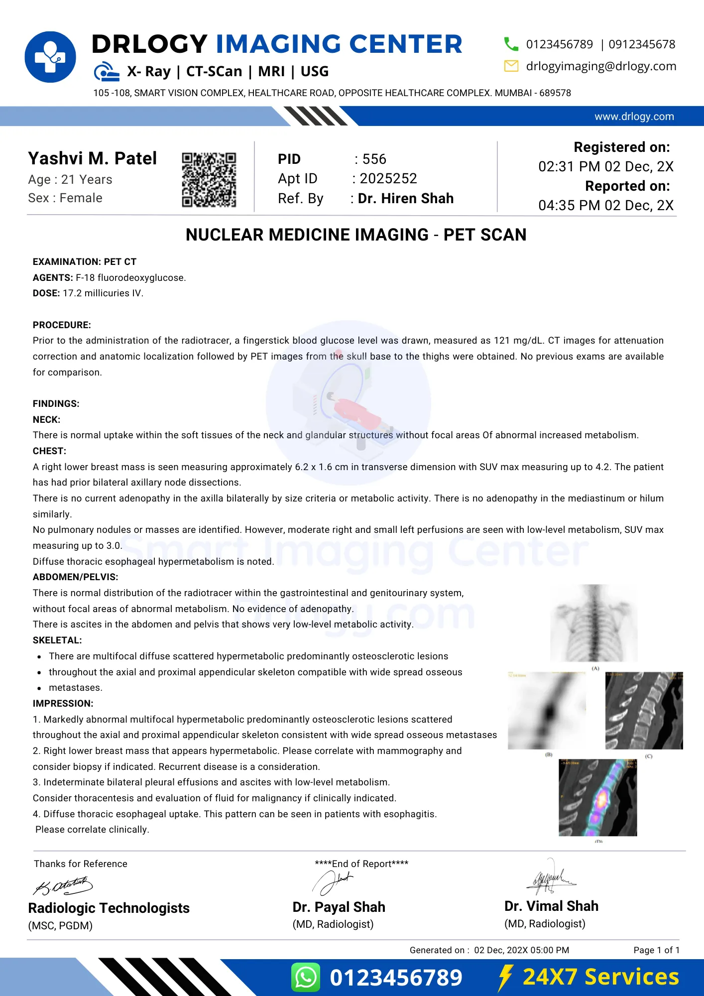
Nuclear Medicine Imaging Report
Nuclear Medicine Imaging Limitation
Here are some limitation associated with a Nuclear Medicine Imaging.
- Radiation exposure
- Limited anatomical detail
- Limited availability of radiotracers
- Longer imaging times
- Potential allergic reactions to radiotracers
Nuclear Medicine Imaging Risk Factors
Here are some risk factors associated with a Nuclear Medicine Imaging
- Exposure to ionizing radiation
- Small amount of radioactive material used
- Potential for allergic reactions to radiotracers
- Minimal discomfort during injection
- Risks associated with specific radiotracers used
- Operator expertise crucial for safety
- Special precautions for pregnant or breastfeeding individuals
Exploring the Safety of Nuclear Medicine Imaging: Myth vs Reality
| Myth | Reality |
|---|---|
| High radiation risk | Low radiation exposure |
| Dangerous for all | Specific medical conditions |
| Harmful for all | Risk factors considered |
| Causes side effects | Generally well-tolerated |
| Permanent effects | Temporary radiation |
| Not for children | Used in pediatric medicine |
| Always invasive | Varies by procedure |
Nuclear Medicine Imaging Price
Here are the estimated Nuclear Medicine Imaging Price in India with different top cities:
| City | Price Range (INR)* |
|---|---|
| Mumbai | 450 - 900 |
| New Delhi | 500 - 900 |
| Bangalore | 450 - 900 |
| Hyderabad | 500 - 900 |
| Kolkata | 450 - 900 |
| Pune | 500 - 900 |
| Lucknow | 450 - 900 |
| Noida | 500 - 900 |
| Surat | 500 - 900 |
| Gurugram | 450 - 900 |
| Patna | 450 - 900 |
| Chennai | 500 - 900 |
| Jaipur | 500 - 900 |
| Ahmedabad | 450 - 900 |
*Prices are approximate and range may vary as per location, facility, type, and procedure.
Summary
Overall, Nuclear Medicine Imaging offers valuable insights into medical conditions using small amounts of radioactive material and is generally safe when managed by trained professionals. Also check Drlogy Test for detailed information about all medical tests for patients, doctors, scholers and medical students.
Reference


