Mammography (Mammogram) For Breast Cancer
Mammography, often called a mammogram, is a breast cancer screening and diagnostic tool that uses low-dose X-rays to create detailed images of the breast tissue, helping detect and diagnose breast cancer at an early stage.
What is Mammography
Mammography is a type of medical imaging.
- It focuses on examining breast tissue.
- It uses X-ray technology to create breast images.
- Often used for breast cancer screening.
- Helps detect tumors and abnormalities.
- Essential for early cancer diagnosis.
- Commonly performed on women as a preventive measure.
Mammography
Here are the basic details for the Mammography.
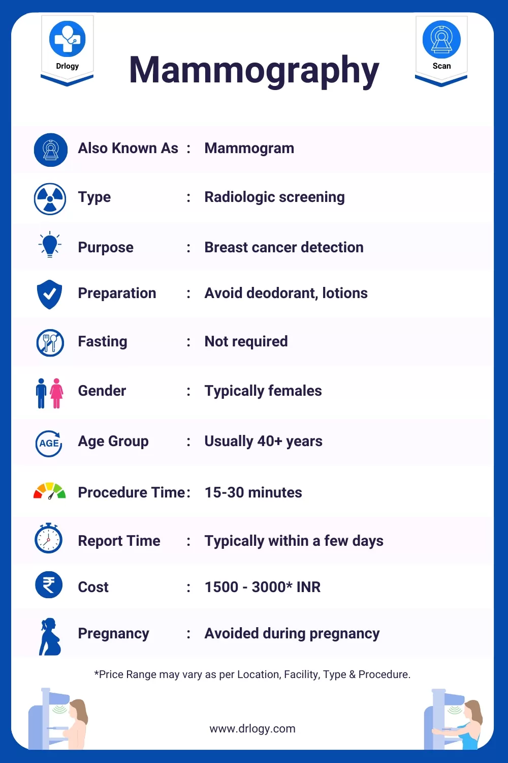
| Also Known As | Mammogram |
| Type | Radiologic screening |
| Purpose | Breast cancer detection |
| Preparation | Avoid deodorant, lotions |
| Fasting | Not required |
| Gender | Typically females |
| Age Group | Usually 40+ years |
| Procedure Duration | 15-30 minutes |
| Reporting Time | Typically within a few days |
| Cost | 1500 - 3000* INR |
| Pregnancy Consideration | Avoided during pregnancy |
| Risks and Safety | Low radiation exposure, generally safe |
| Accessibility | Available in medical centers |
*Price range may vary as per location, facility, type, and procedure.
What are the Purpose or Reasons for Mammography?
Here are common reasons for Mammography.
- Screen for early breast cancer detection
- Detect breast abnormalities or tumors
- Monitor breast health and changes over time
- Guide further diagnostic procedures if needed
- Improve chances of successful treatment for breast cancer

Types of Mammography
Here are 2 main types of Mammography along with their primary use.
| Mammography Type | Organ/System | Primary Use |
|---|---|---|
| Digital Mammography | Breasts | Detect and screen for breast cancer |
| 3D Mammography (Tomosynthesis) | Breasts | Improved breast cancer detection |
These mammography types focus on breast cancer screening and detection.
Preparing for Your Mammography: Tips and Information
Here is the basic preparation before, during, and after Mammography for any patient.
Before the Mammography:
- Consultation: Schedule the mammography and discuss any concerns or medical history with your healthcare provider.
- Fasting: Generally, fasting is not required, but follow any specific instructions provided by your healthcare team.
- Medications: Inform your healthcare provider about any medications you are taking. You may need to avoid certain deodorants, lotions, or powders on the day of the mammogram.
- Clothing: Wear a two-piece outfit on the day of the exam for ease of undressing from the waist up.
- Jewellery and Accessories: Remove jewellery and accessories from the chest area to ensure clear images.
During the Mammography:
- Positioning: You will be positioned in front of the mammography machine, and each breast will be compressed between two plates for imaging.
- Compression: Compression is applied to obtain the best possible images. It may feel uncomfortable but is usually brief.
- Communication: You can communicate with the technician if you have any concerns or discomfort during the procedure.
After the Mammography:
- Recovery: There is typically no special recovery needed. You can resume your normal activities immediately.
- Results: Your mammography results will be reviewed by a radiologist, and a report will be sent to your healthcare provider.
- Follow-Up: Schedule a follow-up appointment with your healthcare provider to discuss the mammography results and any further steps, if necessary.
Remember that specific instructions may vary depending on your individual case and the protocols of the healthcare facility. Always follow the guidance provided by your healthcare team for a successful and safe mammogram.
Who Performs a Mammography?
| Professional | Role |
|---|---|
| Radiologic Technologist | Positions and operates the machine, and assists with imaging. |
| Radiologist | Interprets the mammogram images, and provides a report. |
Mammography Procedure
The procedure for Mammography typically follows these steps:
- Check-in and registration at the clinic/hospital.
- Change into a gown provided by the facility.
- You'll stand in front of the mammography machine.
- A radiologic technologist positions your breast on a platform.
- A clear plastic paddle is gently pressed against your breast to flatten it.
- The machine takes X-ray images from different angles.
- This process is repeated for the other breast.
- You may feel slight discomfort during compression.
- The entire procedure usually takes around 20 minutes.
- The images are reviewed by a radiologist for any abnormalities.
- You'll be informed of the results, which may require further testing if necessary.
Mammography Results
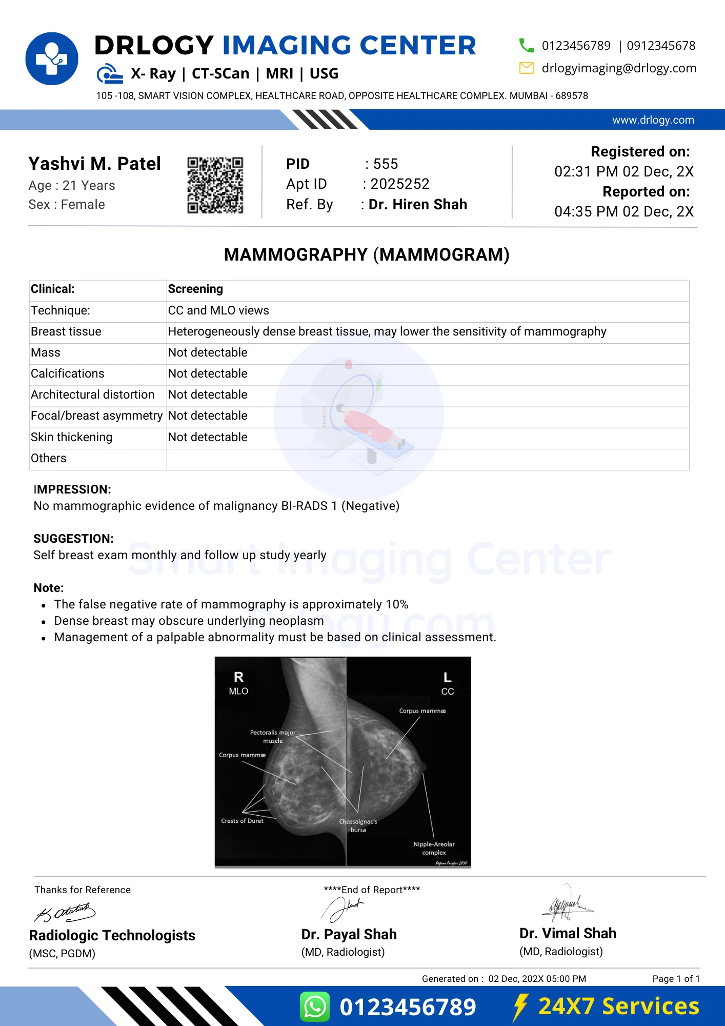
Here are some common elements you might find in a Mammography report:
| Mammography Findings | Interpretation |
|---|---|
| Breast Density | Categories: Almost entirely fatty, scattered fibroglandular tissue, heterogeneously dense, extremely dense |
| Masses or Lumps | Location, size, shape, and characteristics of any detected masses |
| Calcifications | Description of any calcifications, such as microcalcifications |
| Architectural Distortions | Presence or absence of distortions in breast tissue |
| Asymmetries | Any observed asymmetries in breast tissue |
| Skin or Nipple Changes | Noting any skin or nipple abnormalities |
| Axillary Lymph Nodes | Evaluation of axillary lymph nodes for enlargement or abnormalities |
| Impression | Summary of key findings |
| Recommendations | Follow-up tests, additional imaging, or biopsy if necessary |
| Conclusion | Final remarks or clinical recommendations |
It's important to remember that mammography results are interpreted by radiologists or healthcare professionals, and any abnormal findings should be discussed with a healthcare provider for further evaluation and guidance.
Mammography Abnormal Results
Here's a simplified potential causes of abnormal mammography results:
| Abnormal Mammography Finding | Potential Causes |
|---|---|
| Mass or lump | Benign tumor, cyst, cancer |
| Microcalcifications | Ductal carcinoma in situ (DCIS), benign calcifications |
| Architectural distortion | Cancer, scarring, post-surgical changes |
| Asymmetry | Benign breast tissue, positional differences |
| Skin or nipple changes | Skin infection, cancer, benign skin conditions |
| Axillary lymph node enlargement | Infection, inflammation, cancer spreading to lymph nodes |
Abnormal mammography findings should always be evaluated by a healthcare provider, typically with follow-up tests or a biopsy to determine the underlying cause and appropriate next steps.
How Long Does a Mammography Take?
The duration of mammography can vary, but here's a general guideline for the different stages of the procedure:
| Stage of Mammography | Time |
|---|---|
| Registration and paperwork | 5-10 minutes |
| Changing into a gown | 2-5 minutes |
| Imaging process (per breast) | 5-10 minutes |
| Waiting for images to be reviewed | 5-10 minutes |
| Optional additional views | 5-10 minutes (per view) |
| Total Time (for both breasts) | Approximately 20-45 minutes |
Please note that these times are approximate and can vary depending on factors like the facility's efficiency and the need for additional views or procedures. It's a good practice to arrive a little early for your appointment to allow time for registration and paperwork.
Mammography Report

Mammography Limitation
Here are some limitations associated with Mammography.
- Radiation exposure
- Dense breast tissue
- False positives/negatives
- Not for all ages
- Interpretation errors
Mammography Risk Factors
Here are some risk factors associated with Mammography
- Age
- Family history
- Genetic mutations (BRCA)
- Hormone therapy
- Radiation exposure
- Previous breast cancer
- Dense breast tissue
Exploring the Safety of Mammography: Myth vs Reality
| Myth | Reality |
|---|---|
| Causes cancer | Low radiation risk |
| Always detects cancer | Misses some cancers |
| No risk factors for false results | False positives and negatives |
| Unsafe during pregnancy | Safe during pregnancy |
Mammography Price
Here are the estimated Mammography Prices in India with different top cities:
| City | Price Range (INR)* |
|---|---|
| Mumbai | 1500 - 3000 |
| New Delhi | 1800 - 3000 |
| Bangalore | 1500 - 3000 |
| Hyderabad | 1800 - 3000 |
| Kolkata | 1500 - 3000 |
| Pune | 1800 - 3000 |
| Lucknow | 1500 - 3000 |
| Noida | 1800 - 3000 |
| Surat | 1800 - 3000 |
| Gurugram | 1500 - 3000 |
| Patna | 1500 - 3000 |
| Chennai | 1800 - 3000 |
| Jaipur | 1800 - 3000 |
| Ahmedabad | 1500 - 3000 |
*Prices are approximate and the range may vary as per location, facility, type, and procedure.
Summary
Overall, Mammography is a valuable tool for breast cancer screening, despite its limitations and misconceptions. Also check Drlogy Test for detailed information about all medical tests for patients, doctors, scholers and medical students.
Reference


