
Brain MRI: Purpose, Preparation, Procedure, Results & Price
What is a Brain MRI?
A Brain MRI (Magnetic Resonance Imaging) is a non-invasive medical imaging technique that uses strong magnetic fields and radio waves to create detailed images of the structures inside the brain. It is a valuable diagnostic tool used by healthcare professionals to visualize the brain and its various components, including the brain tissue, blood vessels, and any abnormalities or pathology that may be present.
At present, MRI stands out as the most effective imaging method for examining your head, especially your brain, when compared to alternative imaging techniques like CT (computed tomography) scans or X-rays.
Brain MRI Scan
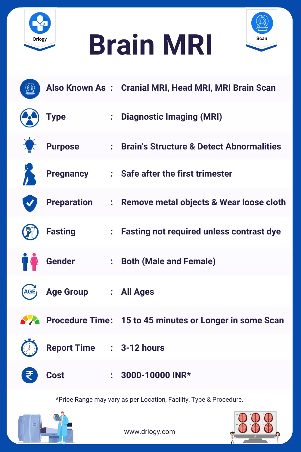
Here are the basic details for the MRI Brain.
| Also Known As | Cranial MRI, Head MRI, MRI Brain Scan |
| Type | Diagnostic Imaging |
| Purpose | To visualize and assess the brain's structure, detect abnormalities, and aid in diagnosis and treatment planning for various neurological conditions. |
| Sample Type | No sample required; non-invasive imaging procedure. |
| Preparation | - Remove metal objects (e.g., jewelry, hairpins) - Wear comfortable, metal-free clothing - Inform the healthcare team about metal implants or devices - Follow any specific instructions from the healthcare provider. |
| Fasting | Typically, fasting is not required unless contrast dye (gadolinium) is used. |
| Gender | All Genders. |
| Age-Group | All Ages |
| Procedure Duration | 15 to 45 minutes, depending on the type and complexity of the scan. More advanced or specialized scans may take longer. |
| Report Time | 3-12 hours |
| Cost | 3000 - 10000 INR* |
| Claustrophobia Options | Open MRI machines or sedation options are available for individuals with claustrophobia or anxiety to make the experience more comfortable. |
| Contrast Dye Use | Contrast dye (gadolinium) may be used to enhance image clarity. It's administered through an IV injection during the scan. |
| Pregnancy Considerations | Brain MRIs are generally considered safe during pregnancy, especially after the first trimester. |
| Risks and Safety | Brain MRIs are non-invasive and typically safe. However, there are potential risks associated with contrast dye use, especially for individuals with kidney issues. |
| Follow-Up and Consultation | Your healthcare provider will discuss the MRI results with you, providing insights into any findings and potential next steps for diagnosis or treatment. |
| Accessibility | Brain MRI services are available at various healthcare facilities, hospitals and imaging centers. |
*Price range may vary as per location, facility, type, and procedure.
Why do I need a Brain MRI?
Headaches: If you experience severe or recurring headaches, your doctor may order a Brain MRI to rule out underlying causes such as tumors, vascular abnormalities, or other structural issues.
Neurological Symptoms: If you have unexplained neurological symptoms like seizures, weakness, numbness, tingling, difficulty speaking, or changes in vision, an MRI can helpentify any abnormalities in the brain that may be causing these symptoms.
Trauma: Following a head injury, especially if it's severe or associated with loss of consciousness, a Brain MRI can help assess for any brain damage, bleeding, or swelling.
Tumors: MRI is a valuable tool for detecting and characterizing brain tumors. It can help determine the location, size, and characteristics of tumors in the brain.
Vascular Abnormalities: MRI can reveal vascular abnormalities in the brain, such as aneurysms, arteriovenous malformations (AVMs), or blocked blood vessels. Detecting these issues early is crucial for treatment.
Monitoring Existing Conditions: For patients with known brain conditions or tumors, regular MRI scans may be recommended to monitor the progress of the condition, the effectiveness of treatment, or to check for any recurrence.
Infections or Inflammatory Conditions: MRI canentify infections, inflammation, or autoimmune diseases affecting the brain, such as multiple sclerosis.
Psychiatric Evaluation: In some cases, Brain MRI may be used to assist in the evaluation of certain psychiatric conditions to rule out any organic causes.
Research and Clinical Trials: Brain MRI is also used in research and clinical trials to study various neurological conditions, which can provide insights into new treatments and therapies.
Preparing for Your MRI Brain Scan: Tips and Information
Preparing for Your MRI Brain Scan involves steps to ensure the procedure goes smoothly and yields accurate results.
Here's what you should do before, during, and after the MRI:
Before the MRI:
Consult with Your Healthcare Provider: Discuss your medical history, any allergies, and inform your healthcare provider if you have any metal implants or devices in your body (e.g., pacemaker, cochlear implant) that might interfere with the MRI.
Dress Comfortably: Wear loose-fitting, comfortable clothing without metal zippers or buttons. In some cases, you may be asked to change into a hospital gown.
Remove Metal Items: Remove all metal objects, including jewelry, watches, eyeglasses, and hairpins. Some MRI facilities may require you to leave these items at home or in a secure locker.
Medications: Follow your doctor's instructions regarding any medications you are taking. You may be asked to temporarily stop taking certain medications, particularly those that affect blood clotting.
Fasting: If contrast dye (gadolinium) will be used, your healthcare provider may advise you not to eat or drink for a few hours before the scan.
Relaxation Techniques: If you're anxious about the MRI, consider relaxation techniques such as deep breathing or meditation to help you stay calm during the procedure.
During the MRI:
Positioning: You'll be positioned on a padded table, and a radiologic technologist will help you get comfortable. It's crucial to lie still during the scan to obtain clear images.
Communication: You'll be given a call button to communicate with the technologist throughout the procedure in case you have any concerns or discomfort.
Noise: MRIs can be noisy due to the magnetic fields. You may be offered earplugs or headphones to minimize the noise.
Contrast Dye: If contrast dye is needed, it will be injected into a vein in your arm during the scan. You may feel a warm sensation or metallic taste, which is normal.
Duration: The MRI may take anywhere from 15 minutes to an hour or more, depending on the type of scan and the information needed.
After the MRI:
Resuming Normal Activities: In most cases, you can resume your regular activities immediately after the MRI. There are typically no restrictions on eating, drinking, or driving.
Review Results: Your MRI images will be reviewed by a radiologist, and the results will be shared with your healthcare provider. They will discuss the findings and any further steps or treatments if necessary.
Follow-Up: Follow any post-scan instructions provided by your healthcare provider. If you experience any unusual symptoms after the MRI, contact your healthcare team promptly.
Remember that MRI Brain Scans are safe and non-invasive, and proper preparation can help ensure a successful and comfortable experience. If you have any questions or concerns about your MRI, don't hesitate to discuss them with your healthcare provider or the MRI technologist before the procedure.
Who Performs a Brain MRI?
A brain MRI is typically performed by a trained team of healthcare professionals. The MRI technologist operates the MRI machine, ensuring proper patient positioning and image quality. Radiologists, who are specialized medical doctors, interpret the MRI images to make diagnoses. Nurses or technicians may assist with patient preparation and contrast dye administration when needed. This collaborative approach ensures a safe and effective brain MRI examination.
Brain MRI Procedure
Here are the 7 steps involved in a typical Brain MRI procedure:
Patient Preparation: Before the MRI, the patient is informed about the procedure and any specific instructions. This may include removing metal objects like jewelry, wearing comfortable, metal-free clothing, and disclosing any metal implants or devices in the body.
Positioning: The patient is positioned on a padded table, usually lying flat on their back. Proper positioning is crucial to ensure clear imaging of the brain.
Safety Screening: The MRI technologist conducts a safety screening to confirm that there are no metallic objects on the patient's body that could be attracted to the strong magnetic field of the MRI machine. If necessary, the patient may be asked to change into a hospital gown.
Injection of Contrast Dye (Optional): If the MRI requires contrast enhancement for better visualization, the technologist or nurse will insert an intravenous (IV) line into a vein in the patient's arm. Contrast dye (usually gadolinium) is then injected through the IV during the scan.
Entering the MRI Machine: The patient is moved into the MRI machine, which is a large, cylindrical device with a central opening. The patient's head is typically placed inside a specialized coil designed for brain imaging. It's essential to remain as still as possible during the scan to avoid blurring the images.
Image Acquisition: The MRI technologist operates the machine from a separate control room but maintains communication with the patient throughout the procedure. The MRI machine generates powerful magnetic fields and radio waves to create detailed cross-sectional images of the brain. Various sequences and angles may be used to capture the necessary images.
Completing the Procedure: Once all the required images are obtained, the MRI technologist will exit the room and the patient is usually moved out of the machine. If contrast dye was used, the IV line may be removed. The patient can then leave the imaging facility.
The MRI images are later reviewed by a radiologist, who interprets the results and provides a report to the referring healthcare provider. This report is used for diagnosis and treatment planning.
MRI Brain Scan Results
MRI Brain scan results typically consist of a detailed report generated by a radiologist who has reviewed the images obtained during the scan.
Here's what you can expect from MRI Brain scan results:
Technical Information: The report usually begins with technical details about the MRI scan, including the date, time, and location of the scan, as well as the specific type of MRI performed (e.g., T1-weighted, T2-weighted, with or without contrast).
Clinical Indications: The report may include information about the reason for the MRI, often referred to as clinical indications. This section explains why the scan was ordered and what symptoms or medical concerns led to the decision.
Image Findings: The core of the report details the radiologist's findings based on the MRI images. This section describes the appearance of various brain structures and notes any abnormalities or significant findings. Common findings may include the presence of tumors, vascular abnormalities, areas of inflammation, or other structural issues.
Impressions and Recommendations: The radiologist provides their impressions and conclusions based on the MRI findings. They may offer a preliminary diagnosis or suggest further evaluation, additional tests, or consultations with specialists if necessary. Recommendations for treatment or management may also be included.
Comparison with Previous Scans: If available, the report may compare the current MRI findings with previous scans to track changes over time. This can be valuable for assessing the progression of certain conditions.
Clinical Correlation: The radiologist may suggest that the MRI findings need to be correlated with the patient's clinical history, symptoms, and other medical information. This emphasizes the importance of a collaborative approach between the radiologist and the referring healthcare provider.
Limitations: The report may acknowledge any limitations of the MRI study, such as factors that could have affected image quality or areas that were challenging to assess due to motion artifacts or other issues.
Final Impressions and Signature: The report typically concludes with the radiologist's final impressions, their name, and a signature, confirming the accuracy and reliability of the findings.
It's important to note that MRI Brain scan results are intended for healthcare professionals and are part of the diagnostic process. The referring healthcare provider will discuss the results with the patient, explain their significance, and develop a treatment plan if needed.
How Long Does a Brain MRI Take?
The duration of a Brain MRI (Magnetic Resonance Imaging) can vary depending on several factors, including the type of scan being performed, the specific protocols used, and the patient's cooperation.
Here are some general guidelines:
| Factor | Typical Duration |
|---|---|
| Routine Brain MRI | 15 - 30 minutes |
| Brain MRI with Contrast | 30 - 45 minutes |
| Advanced or Specialized Scans | 45 minutes to 1+ hour |
| Patient Factors (e.g., breaks) | Variable |
| Facility and Equipment | Variable |
MRI Brain Report

Brain MRI Limitation
Here are three common limitations of Brain MRI:
Motion Artifacts: Patient movement during the scan can lead to blurred images and reduce diagnostic accuracy.
Metallic Implants: Metal objects or implants in the body can cause distortion in MRI images, making it challenging to visualize certain areas.
Limited Functional Information: While MRI provides excellent structural details, it may not capture real-time functional changes in the brain, unlike techniques like fMRI (functional MRI).
Brain MRI Risk Factors
- No Ionizing Radiation: Brain MRIs do not involve ionizing radiation, reducing the risk of radiation-related harm.
- Contrast Dye Risks: In rare cases, contrast dye used in MRIs may cause allergic reactions or complications, especially in individuals with kidney issues.
- Metallic Implants: Certain metal implants or devices in the body can pose risks during MRI, potentially causing discomfort or displacement.
Exploring the Safety of MRI Brain Scans: Myth vs. Reality
Here highlights some common misconceptions about MRI brain scans and provides accurate information to address these myths, emphasizing the safety and effectiveness of MRI as a diagnostic tool.
| Myths | Realities |
|---|---|
| MRI scans expose you to harmful radiation. | MRI does not use ionizing radiation (X-rays), making it safer than CT scans in terms of radiation exposure. It relies on strong magnetic fields and radio waves to create images. |
| Having metal in your body is dangerous during an MRI. | While certain metal implants can pose risks, many implants are MRI-safe. Technological advancements and careful screening help ensure patient safety during MRI scans. |
| MRI scans can erase data from credit cards and electronic devices. | MRI's magnetic fields can affect magnetic stripe cards, but the risk to modern credit cards is minimal. It won't damage or erase data from smartphones or other electronic devices. |
| MRI scans are claustrophobic and uncomfortable. | MRI machines can be tight and noisy, which may cause discomfort for some individuals. However, open MRI machines or techniques to reduce noise and anxiety can make the experience more comfortable. |
| The contrast dye used in MRI scans is harmful. | Gadolinium-based contrast agents are generally safe but may have rare side effects. Healthcare providers carefully consider the need for contrast and monitor patients for adverse reactions. |
| MRI scans take a long time to complete. | The duration of an MRI scan varies, but most routine brain scans take around 15-30 minutes. Longer scans may be necessary for more detailed imaging. |
| MRI scans are not suitable for individuals with tattoos. | While some older tattoos may contain metallic ink, modern tattoos typically do not pose a problem during MRI scans. Tattoos are unlikely to cause heating or other safety concerns. |
| MRI scans are not safe during pregnancy. | MRI is generally considered safe during pregnancy, especially after the first trimester. However, healthcare providers carefully weigh the risks and benefits when recommending MRI during pregnancy. |
Brain MRI Price
Here are the estimated Brain MRI Scan Price in India with different top cities:
| City | Price Range (INR)* |
|---|---|
| Mumbai | 4000 - 8000 |
| New Delhi | 4000 - 8000 |
| Bangalore | 5000 - 10000 |
| Hyderabad | 5000 - 8000 |
| Kolkata | 3500 - 6000 |
| Pune | 4000 - 7000 |
| Lucknow | 4000 - 7000 |
| Noida | 4000 - 8000 |
| Surat | 4000 - 7000 |
| Gurugram | 4000 - 6000 |
| Patna | 3000 - 5500 |
| Chennai | 4000 - 10000 |
| Jaipur | 4000 - 8000 |
| Ahmedabad | 4500 - 7000 |
*Prices are approximate and range may vary as per location, facility, type, and procedure.
Reference
- Magnetic resonance imaging of the brain - Wikipedia [1].
- Magnetic Resonance Imaging of the Living Brain - NCBI [2].
FAQ
How long does MRI of Brain take?
The duration of a Brain MRI (Magnetic Resonance Imaging) can vary depending on factors such as the type of scan, the complexity of the images needed, and the individual patient. However, as a general guideline:
- A routine Brain MRI without contrast typically takes approximately 15 to 30 minutes.
- If contrast dye (gadolinium) is used to enhance the images, the scan may take a bit longer, typically around 30 to 45 minutes.
Why would a Doctor order an MRI of the Brain?
A doctor may order an MRI (Magnetic Resonance Imaging) of the brain for a variety of medical reasons, including:
- Detection of a blood clot in the brain.
- Assessment of a brain aneurysm.
- Evaluation of a brain hemorrhage.
- Diagnosis and monitoring of brain infections like encephalitis.
- Examination of brain damage associated with epilepsy.
- Detection and characterization of brain tumors and cysts.
- Evaluation of chronic neurological conditions, such as multiple sclerosis (MS).
- Assessment of dementia and related cognitive disorders.
- Investigation of hydrocephalus.
- Diagnosis of pituitary gland issues, such as pituitary adenoma.
- Detection and evaluation of stroke.
- Assessment of brain development or structural issues, including Chiari malformation and malformations of cortical development.
- Diagnosis and monitoring of traumatic brain injuries (TBI).
Which is better MRI or CT Scan for brain?
Whether an MRI (Magnetic Resonance Imaging) or a CT (Computed Tomography) scan is better for evaluating the brain depends on the specific clinical situation and what the healthcare provider is trying to assess.
MRI for Brain:
Advantages:
- Provides highly detailed images of soft tissues, making it excellent for detecting small abnormalities and subtle changes in the brain.
- Does not use ionizing radiation, which is advantageous for pregnant women and repeated imaging.
- Can capture images in multiple planes (e.g., axial, sagittal, coronal) without repositioning the patient.
- Ideal for assessing neurological conditions, tumors, multiple sclerosis, and soft tissue abnormalities.
Limitations:
- MRI is generally more time-consuming than CT scans, which can be a concern for patients with claustrophobia or those who cannot remain still for an extended period.
- Some patients may not be suitable candidates for MRI due to metallic implants or claustrophobia.
- Cost is typically higher than CT scans.
CT Scan for Brain:
Advantages:
- Extremely fast imaging, making it suitable for patients who cannot tolerate lengthy scans.
- Excellent for detecting acute bleeding in the brain (e.g., after head trauma or stroke).
- Can be performed on patients with metallic implants (although there may still be some limitations).
Limitations:
- Uses ionizing radiation, which can be a concern for repeated imaging or pregnant women.
- Provides less detailed images of soft tissues compared to MRI.
- May not be as effective in detecting small tumors or subtle abnormalities in the brain.
The choice between MRI and CT for brain imaging depends on the clinical context and the specific diagnostic needs. In cases where a high level of detail is required to assess soft tissue structures or when evaluating chronic neurological conditions, an MRI is typically preferred.
However, for acute situations like head trauma or suspected bleeding in the brain, a CT scan's speed and ability to detect acute abnormalities can be advantageous. The healthcare provider will consider these factors when ordering the appropriate imaging study.


